Highlights
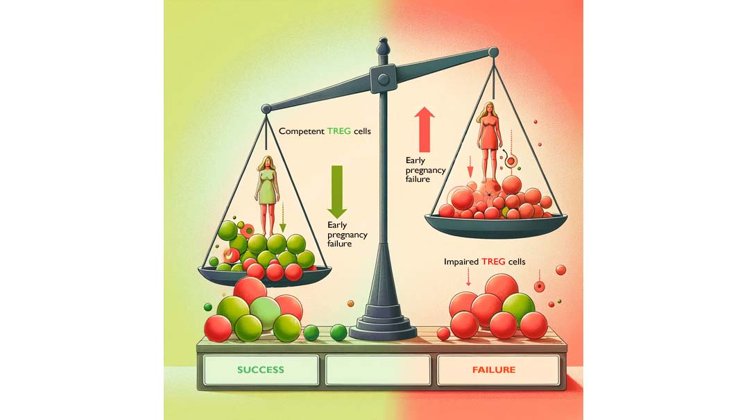
A recent study published by Dr Robertson’s group reveals that women experiencing recurrent implantation failure and/or recurrent pregnancy losses have, prior to conception:
- fewer CD4+ regulatory T cells in the peripheral blood.
- these Treg cells have an altered phenotype with:
- reduced suppressive competence
- a pro-inflammatory shift
- similarities with autoimmune conditions
Regulatory T cells (Treg cells) are key specialized immune cells that promote maternal-fetal tolerance1-2:
- by suppressing inflammation
- by preventing the uterus from rejecting the embryo through the secretion of factors suppressing the maternal immune response to “foreign” paternal components called antigens.
- are involved in the uterine spiral artery remodeling required for placentation.
Treg cells can arise from the blood and be recruited within the decidua3 where they are “primed” by sex hormones and factors in the seminal fluid then sequestrated by trophoblast secretion4, or they may be generated locally (within the uterus) activated by the presence of the embryo (HLA-G expressed on the surface of the trophoblast)5. Lower Treg levels have been reported in the uterus of women with recurrent implantation failures, recurrent miscarriage6 and pregnancy complications such as pre-eclampsia.
In this study, the authors compared systemic (in peripheral blood samples) Treg cell profile of women with early pregnancy failures to women with proven fertility.
Results show that Treg cells are fewer and have an altered phenotype in early pregnancy failure (EPF) women.
- Treg cells number was 32% lower in EPF women which may be linked to a higher rate of maturation leading to Treg cell exhaustion in the peripheral blood
- These cells had a 20% lower expression of Foxp3 (a specific marker of Treg cells)
- Treg cells were 34% less stable
- Treg cells had 21% lower suppressive capacity
- this is coupled with increased transcription of pro-inflammatory cytokine (IL-17, TNF-α, IL17A)
These changes may disrupt pregnancy tolerance and impair embryo implantation as the majority of decidual T cells originate from the peripheral blood.
Regulatory T cell assessment in the peripheral, prior to conception, may help determine the possible causes of your implantation failure or pregnancy losses.
Some immune therapies may help increase Treg cells expansion and recruitment to the decidua.
References
- Erlebacher, A. (2013). Mechanisms of T cell tolerance towards the allogeneic fetus. Nat. Rev. Immunol. 13, 23–33.
- Robertson, S.A., Care, A.S., and Moldenhauer, L.M. (2018). Regulatory T cells in embryo implantation and the immune response to pregnancy. J. Clin. Invest. 128, 4224–4235.
- Tilburgs T, Roelen DL, van der Mast BJ, de Groot-Swings GM, Kleijburg C, Scherjon SA, Claas FH. Evidence for a selective migration of fetus-specific CD4+CD25bright regulatory T cells from the peripheral blood to the decidua in human pregnancy. J Immunol. 2008 Apr 15;180(8):5737-45.
- Schumacher A, Brachwitz N, Sohr S, Engeland K, Langwisch S, Dolaptchieva M, Alexander T, Taran A, Malfertheiner SF, Costa SD, Zimmermann G, Nitschke C, Volk HD, Alexander H, Gunzer M, Zenclussen AC. Human chorionic gonadotropin attracts regulatory T cells into the fetal-maternal interface during early human pregnancy. J Immunol. 2009 May 1;182(9):5488-97.
- Tilburgs, T., Crespo, Á.C., van der Zwan, A., Rybalov, B., Raj, T., Stranger, B., Gardner, L., Moffett, A., and Strominger, J.L. (2015). Human HLA-G+ extravillous trophoblasts: Immune-activating cells that interact with decidual leukocytes. Proc. Natl. Acad. Sci. USA 112, 7219–7224.
- Granne, I., Shen, M., Rodriguez-Caro, H., Chadha, G., O’Donnell, E., Brosens, J.J., Quenby, S., Child, T., and Southcombe, J.H. (2022). Characterisation of peri-implantation endometrial Treg and identification of an altered phenotype in recurrent pregnancy loss. Mucosal Immunol. 15, 120–129.
More to read
- Research Leptin and Pregnancy: Research Review
- Research Thyroid Function and Pregnancy: Research Review
- Research PAI-1, Thrombophilia and Pregnancy: Research Review
- Research HLA Antibodies and Pregnancy: Research Review
- Research Blood Clotting and Pregnancy
- Research Natural Killer Cells and Pregnancy: Research Review